The short answer is: It is a method for 3D printing skeletons of living animals as shown recently by Notre Dame students and a rep from MakerBot.
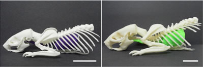
3d printing possibilities continue to fascinate people around the world. Looks like this is just one of the myrind of other applications bound to come up in days to come. Innovations with 3d printers have just about started. The researchers have outlined a method for printing out skeletal structures of live mice, rats and rabbits in plastic using CT scanning.
Traditionally, models have been created using mold-injection methods.This method does have some limitations including the difficulty in making highly complex products in a timely, cost-effective manner.
Three-dimensional printing using as additive manufacturing process allows detailed objects to be 3d printed.
As per the researchers “These printers have the ability to extrude high-resolution objects with enough detail to accurately represent in vivo images
generated from a preclinical X-ray CT scanner.With proper data collection, surface rendering, and stereolithographic editing, it is now possible and inexpensive to rapidly produce detailed skeletal and soft tissue structures from X-ray CT data. ”
You can see further information on this exciting new method at:
http://www.jove.com/video/50250/3d-printing-of-preclinical-x-ray-computed-tomographic-data-sets
This is great news for researchers, students and tutors alike since they can do / show better visualization. A computer screen showing 3d images will not have the same impact.
Another advantage would be that animal anotomies will be better demonstrated without actually dissecting the animal.
Using freeware, engineering student Evan Doney converted data from CT scans to language that is understood by a 3D printer. Then he printed out several PoC skeletons, including a rabbit skull.